Description:
Summary
Vanderbilt researchers have developed bioresorbable RF coils to improve the signal-to-noise ratio (SNR) for use in post-surgical monitoring.
Addressed Need
Current MRI techniques have difficulty imaging certain biomarkers and characteristics internal to the body due to an insufficient signal-to-noise ratio (SNR). One approach to mitigate this barrier is embedding a small MRI coil into the body, increasing the SNR in that specific location. However, using this technique requires the device to either be surgically removed or left within the body, both of which can outweigh the benefits of a higher SNR.
Vanderbilt researchers have developed a technology that eliminates these risks by enlisting the use of bioresorbable materials. This technology can be safely implanted in humans to allow for post-surgical monitoring by MRI with a high SNR value and can image sensitive internal biomarkers, such as nerves, without the risk of harming it.
Technology Description
Our device utilizes small radio frequency coils in order to optimize SNR and develop high resolution MRIs. This bioresorbable inductively-coupled coil (BIC) can be surgically implanted in the body for a period of time (days to weeks) before being harmlessly absorbed into the body. Therefore, the device can accurately measure and monitor a patient for a period of time without the risks of damage and injury during surgical removal.
Commercial Applications
A potential application of the BIC is imaging of the peripheral nerve following surgical repair, but it could also be used in other surgeries where post-surgical monitoring is necessary and unachievable with current technology. The BIC promises a low-cost approach to provide high SNR MRI over a reduced field of view. For post-surgical imaging, the BIC has potential to provide more detailed evaluations of patient’s recovery status, more quickly informing the surgeon if revision surgery is necessary. Ultimately, this device hopes to improve patient outcomes and reduce unnecessary procedures.
Technology Development Status
Preliminary designs have been made. A prototype is in development.
Intellectual Property Status
A patent has been issued: US-2023-0103510-A1

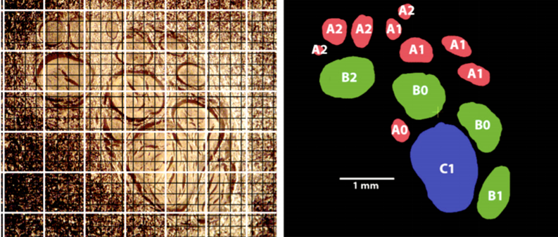
Fig 1: Histologic image of human median nerve (left) and map or segmented fascicles (right). White grid lines show current best in-vivo DTI resolution and black grid lines show possible resolution after 14-fold SNR gain.