Description:
Summary
Vanderbilt researchers have developed a novel brain age estimation framework that leverages diffusion MRI to capture microstructural changes associated with aging. This innovative approach outperforms conventional T1-weighted MRI methods by focusing specifically on subtle brain changes, providing a powerful tool for early detection of neurodegenerative diseases and enabling earlier clinical intervention.
Addressed Need
Current brain age estimation techniques primarily rely on T1-weighted MRI, which fails to capture crucial microstructuralchanges that occur with aging. Early detection of neurodegenerative diseases like Alzheimer's and mild cognitive impairment requires more sensitive biomarkers that can identify subtle changes before symptoms manifest. Additionally, conventional methods often conflate macrostructural and microstructural information, limiting diagnostic precision. This technology directly addresses these challenges by focusing specifically on microstructural signatures of brain aging through advanced diffusion MRI analysis.
Key Benefits
Enhanced diagnostic accuracy for neurodegenerative disease detection compared to conventional methods
Earlier intervention opportunities through identification of advanced brain age before clinical symptoms appear
Improved monitoring of disease progression and treatment efficacy
Potential for personalized treatment strategies based on specific microstructural profiles
Key Features
Novel framework using diffusion MRI to estimate brain age based on microstructural changes
Non-rigid registration technique to minimize macrostructural influence during model training
Superior performance compared to both conventional T1-weighted MRI approaches and state-of-the-art deep learning models
Specialized focus on capturing subtle microstructural signatures of brain aging not apparent in traditional anatomical scans
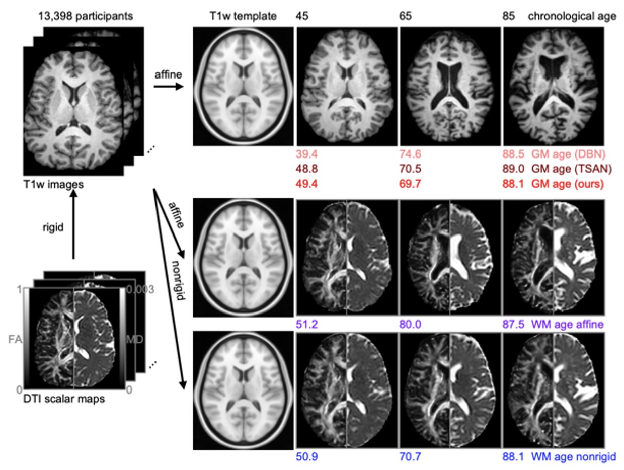
Figure 1
Our proprietary Brain Age Estimation Framework uses diffusion MRI to detect subtle microstructural changes invisible to traditional methods, enabling earlier detection of neurodegeneration and superior diagnostic accuracy compared to conventional approaches—a breakthrough for advancing preventive neurological care.
Intellectual Property Status
Patent application filed.
Stage of Development
The technology has been experimentally validated and demonstrated to outperform conventional T1-weighted MRI-based approaches as well as state-of-the-art deep learning models that use both macro- and microstructural information.
CTTC Contact
Chris Harris – chris.harris@Vanderbilt.Edu
VU Ref. Number
VU25015